3D Medical Radiation Dose Mapping Using Computed Tomography of Fluorescent DNA

Pioneering a new technique for radiation treatment calibration.

Sept 2011 – April 2012
Queen’s University, Kingston
Fourth-Year Thesis Project

3D Medical Radiation Dose Mapping Using Computed Tomography of Fluorescent DNA

Pioneering a new technique for radiation treatment calibration.

Sept 2011 – April 2012
Queen’s University, Kingston
Fourth-Year Thesis Project

My fourth-year undergraduate thesis project focused on biomedical technologies to help calibrate and guide radiation treatment for cancer patients. Under the supervision of the Senior Medical Physicist at Kingston General Hospital, I developed a proof-of-concept simulation and prototype design for creating 3D images of radiation dose distributions using a new technique called DNA Dosimetry.

Precise radiation treatment relies on the careful application of high-energy photons (e.g. x-rays) to kill cancerous cells at the tumor site, while minimizing the dose absorbed by surrounding healthy tissues. The medical linear accelerators (LINACs) used today provide sophisticated control over x-ray beam shape, intensity, and dose angle to optimize the 3D dose pattern delivered to the patient. However, due to the complexity of dose delivery, treatment plans are first tested on non-living targets called “phantoms”, allowing dose discrepancies to be identified and corrected, and thereby providing necessary measures for calibration and quality assurance. 

Fig RadiationTherapy

A modern medical LINAC (top) and patient-tailored treatment plan (bottom) for radiation therapy of tumors.

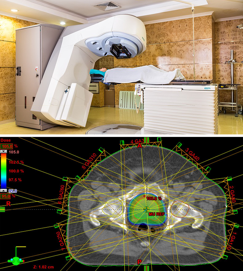
Radiation Dosimetry using DNA Phantoms

3D dosimetry (the process of measuring radiation dose distribution) is typically achieved with a gel-like phantom that turns opaque when exposed to ionizing radiation. However, a more direct measure of biological radiation dose can be obtained by monitoring the fracturing of DNA. This has recently been made possible by the development of “DNA Dosimeters”, consisting of small bio-engineered strands of DNA that, when damaged by the effects of radiation, become fluorescent and therefore emit light when illuminated with the right laser colour. 

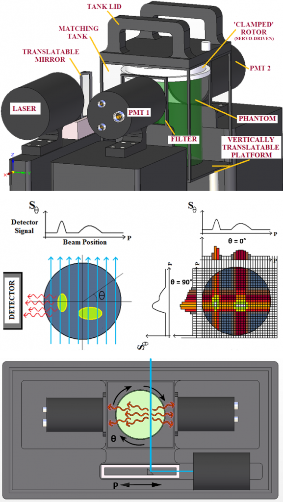
My challenge was to find a way to use this radiation-induced fluorescence to create a 3D reconstruction of the dose distribution, after a jar of the gel-suspended DNA is exposed to the intended radiation treatment plan.

DNA Dosimeter Schematic

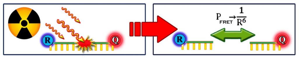
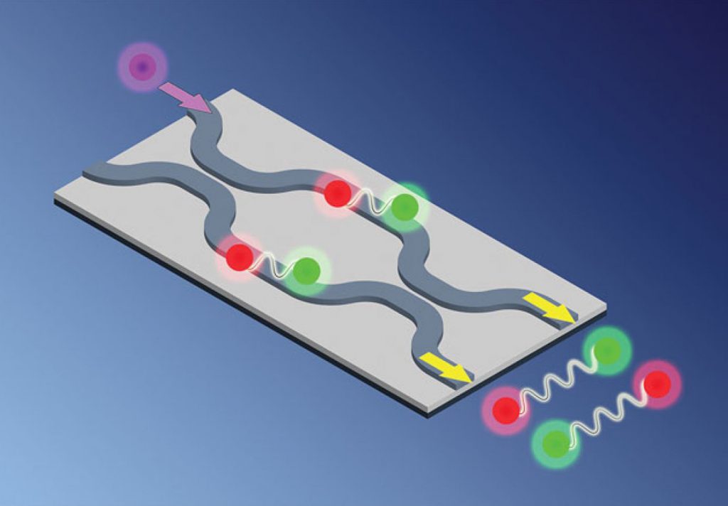
A quencher molecule (Q) suppresses the response of a fluorescent reporter (R) through Förster Resonance Energy Transfer (FRET) until the DNA strand is severed by radiation.

Solution and Prototype

I based my solution on the computed tomography (CT) scans used for patient imaging in a hospital, but rather than measuring the attenuation of a beam through the phantom, I reconstructed 3D images from a collected fluorescence response as a laser beam is scanned through the sample.

Engineering the prototype required design decisions on the pulsed laser system, opto-mechanics and scanning mechanism, signal amplification circuits for the photomultiplier tubes, and the optimal DNA concentration and gel solution for the phantom. The final design selected between several options (such as different photomultiplier tube detection areas) to optimize for scan time, image reconstruction quality, and prototype cost. In addition to scanning the phantom as a function of laser angle-of-incidence and lateral position, the 3D image reconstruction required detailed simulation of the photon scattering physics and optical cross-sections. I also had to design digital filters to de-convolve the reconstructed image from noise and errors caused by scattering and imperfect signal collection.

Fig PrototypeSchematic 3

Outcomes

A simulation of my technique in action!

My completed thesis proved that 3D radiation dose mapping was indeed achievable using DNA dosimeters. The results I obtained showed promise and together with my recommendations for improvements provided a roadmap for the future development of this technology.

The work was extremely well-received, with one of my committee members commenting that it was very close to being a full-fledged Master’s Thesis (just needing a few additional months for prototype assembly and data collection).  

See my full undergraduate thesis here, and my presentation poster here.

Outcomes

My completed thesis proved that 3D radiation dose mapping was indeed achievable using DNA dosimeters. The results I obtained showed promise and together with my recommendations for improvements provided a roadmap for the future development of this technology.

The work was extremely well-received, with one of my committee members commenting that it was very close to being a full-fledged Master’s Thesis (just needing a few additional months for prototype assembly and data collection).  

See my full undergraduate thesis here, and my presentation poster here.

A simulation of my technique in action!

Testimonial from my Supervisor

“The purpose of the thesis course, for which I served as Ryan’s supervisor, is to draw upon and expand one’s entire academic training to solve a (complex) engineering problem. Ryan’s thesis certainly required him to apply his knowledge in many areas. Our weekly 30 minute meetings allowed me, in part, to evaluate his academic strengths and weaknesses. I was extremely impressed with this young man’s knowledge and his ability to gain new knowledge. Based on his written thesis and our interactions, I would say that Ryan was one of the top 2 students I have supervised… I found it remarkable how thorough he was in his discovery phase, how well he articulated his knowledge and questions, and how he put all that knowledge together into an extremely well-written thesis.”

AndrewKerr

Dr. Andrew T. Kerr
Senior Medical Physicist
(Radiation Oncology)
Kingston General Hospital,
Associate Professor (Adjunct)
Queen’s University

Check out some of my other past projects!

I’ve worked on technical projects in a variety of fields. Here are some highlights:

Integrated Photonics

Empowering next-generation optical technologies.

Quantum Computing

My time as a "quantum coder".

Chip-Based Medical Biosensors

Merging engineering with biochemistry.